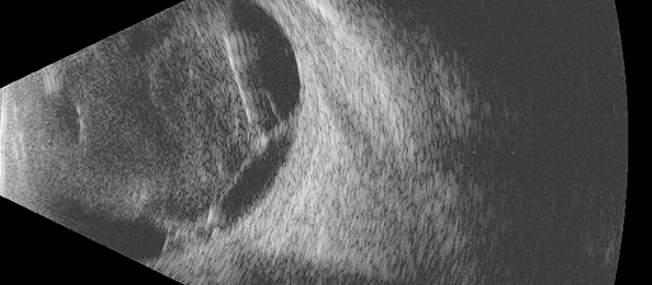
Ophthalmic Ultrasound
When the retina or vitreous cannot be visualized by the lights and lenses used during your eye examination, an ultrasound of the eye may be necessary. This typically occurs when there is too much bleeding inside the eye to allow your doctor to see the retina. Other situations that may require ultrasound include severely advanced cataract or corneal scarring. Ultrasound is also used to diagnose and measure tumors inside the eye.

An ultrasound examination uses sound waves sent from a probe placed on the eye or eyelids to provide “sonar” images of the inside of the eye. The technology is very similar to the kind of ultrasound used to visualize other parts of the body, such as the liver or uterus. The sound waves are painless and harmless to the eye. The images can be printed or stored digitally so that they can be used for comparison on future examinations.
There are various ultrasound probes used for different purposes. These probes are designed to image and measure different parts of the eye. The most commonly used probe is the B-scan probe, which allows the doctor to view the internal structure of the eye through a dense cataract or hemorrhage inside the eye. We also have less commonly used probes, such as the diagnostic A-scan probe and anterior segment high frequency B-scan (also called ultrasound biomicroscopy). These probes are used in our practice primarily for the diagnosis and measurement of tumors inside the eye.


