About Vein Occlusion
The retina gets its nutrients from tiny blood vessels that run through it. In a “vein occlusion,” either the main vein or a smaller tributary vein gets blocked off. When this happens, some bleeding and swelling develop that can make your vision blurry and distorted. Vein occlusions generally happen without a reason, but they are sometimes associated with high blood pressure, clotting problems, or glaucoma.
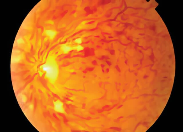
When a central vein occlusion occurs, hemorrhages are present throughout the retina, in all quadrants.
In the early stages of damage, some of the small blood vessels become twisted and small spots of blood appear in the retina. It is important to monitor this stage as it can progress to a more severe form that can cause blindness. After a vein occlusion, these small retinal blood vessels that are normally water-tight start to leak. If the leakage becomes substantial, your vision may become worse. We call this macular edema. To stop the leakage, gentle laser treatment might be done to cauterize the leaky vessels. Sometimes a steroid, anti-vascular endothelial growth factor therapy1 (Ou et al. 2017), or other medication injection is used as well or instead. In many cases these treatments may be combined over time. Treatment can usually help to prevent the vision from worsening and occasionally can make it better. However, not all vein occlusions respond to treatment and in some cases your vision may be permanently damaged. You may require a fluorescein angiogram to assess the blood flow in your eye, which helps us to decide on the appropriate treatment.
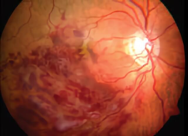
The reduction in blood flow from a branch vein occlusion causes regional bleeding in the retina seen on the left side of this retina photograph.
In the more advanced stages of damage, abnormal blood vessels start to grow on the retina’s surface. These vessels can break and bleed, causing a vitreous hemorrhage. Even worse, these abnormal blood vessels can cause the retina to detach, a potentially blinding problem. In these cases, more aggressive laser treatment is applied to the retina to stop the vessel growth. Laser can prevent blindness. In some cases, the normal retinal blood vessels die off from the vein occlusion (called ischemia) and the vision is permanently damaged.
In very advanced stages of damage, an operation may be needed to clear out the blood in the eye or repair a retinal detachment. This operation, called vitrectomy, involves making three small incisions and using miniature forceps, scissors and other instruments to restore a more normal retinal anatomy. Sometimes, however, the vision cannot be improved despite any efforts.
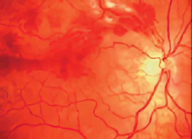
In more severe branch vein occlusions, bleeding may occur from the growth of new abnormal blood vessels into the vitreous cavity in front of the retina.
References
- Ou WC, Brown DM, Payne JF, Wykoff CC. Relationship Between Visual Acuity and Retinal Thickness During Anti-Vascular Endothelial Growth Factor Therapy for Retinal Diseases. Am J Ophthalmol. 2017 Aug;180:8-17. pubmed.ncbi.nlm.nih.gov/28549848
Schedule Vein Occlusion Treatment in Northern California with Retinal Consultants Medical Group
Since 1975, Retinal Consultants Medical Group has been providing outstanding care to patients throughout Northern California, including Sacramento, Modesto, and Stockton. Our retina specialists and surgeons treat multiple vitreoretinal conditions, such as age-related macular degeneration and diabetic retinopathy. We invite you to contact us with any questions or schedule an appointment today.