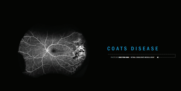
January’s Image of the Month: Coats disease

January’s Image of the Month is of Coats disease. Coats disease is a predominantly unilateral (95%) condition resulting in the progressive development of abnormal retinal vessels. It is typically seen in the temporal retina as telangiectasis with aneurysmal changes that light up like a bulb on angiography. The vascular abnormalities are sometimes seen by themselves, but can also be associated with varying degrees of exudation ranging from focal exudates to total retinal detachment. Pediatric cases can result in leukocoria and be confused with retinoblastoma. When diagnosed in adulthood, as in this case, the pathology tends to be less severe and findings are more likely to be confined to the peripheral retina.


