An Overview of the Choroid and How it Affects Vision

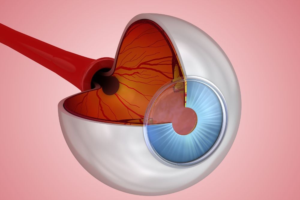
Sandwiched between your sclera and retina lies a multi-layered vascular tissue known as the choroid. This part of the eye is vital to retinal health by providing oxygen and nourishment and regulating temperature. Choroidal disorders, such as bleeding, detachment, or malignant growths, can pose serious risks to vision health. While they can be treated, regular examinations can ensure healthy choroidal function.
The Anatomy of the Choroid
The choroid is part of the uveal tract, which is the middle part of the eye that also includes the iris and ciliary body. The structure of the choroid consists of four layers:
- Haller’s layer: the outermost layer, consisting of larger blood vessels
- Sattler’s layer: a layer of tissue consisting of medium-sized blood vessels
- Choriocapillaries: a layer of tissue consisting of capillaries
- Bruch’s membrane: the innermost layer of the choroid that contains the basement membrane of the retinal pigment epithelium (RPE).
The Function of the Choroid
One of the choroid’s primary functions is to maintain blood circulation in the eye, accounting for about 85% of ocular blood flow. The choroid provides nutrients to the retina, macula, and optic nerve. It also helps regulate the temperature of the retina as well as intraocular pressure.
The choroid is connected to the RPE, a pigmented cell layer that absorbs light and limits damaging reflections within the eye. This pigmentation is due to a molecule called melanin, which absorbs stray light that may interfere with visual images. As melanin also absorbs excess light, it may protect against light toxicity.
Common Disorders of the Choroid
Any choroid conditions or infections may harm the macula and optic nerve, leading to serious vision issues and possibly total blindness.
Choroidal Detachment
Should the choroid move out of placement, due to fluid or blood accumulation, a choroidal detachment can develop. In some cases, there are no symptoms, however, some patients experience soreness in the affected eye or severe pain. Blurriness is also common, although this may depend on related issues, like recent surgery or eye pressure.
In patients who had glaucoma surgery, low eye pressure can occur, leading to serous or hemorrhagic choroidal detachment months or years later. Other risk factors include:
- Use of blood thinners
- Having a short eye (nanophthalmos)
- Eye trauma
- Eye inflammation
- History of choroidal detachment
- Glaucoma
Choroidal Melanoma
Choroidal melanomas are a type of cancer in which malignant tumor cells grow inside the choroid. Although they are rare, they are very serious, as they can cause vision loss and spread to other parts of the body, causing serious health problems and even death. However, with early diagnosis, the chance of survival with appropriate treatment is excellent.
Choroidal melanomas are generally diagnosed by an ophthalmologist during a dilated eye exam. If you have certain risk factors, it’s recommended that you see an ophthalmologist regularly for a comprehensive eye exam in order to catch ocular melanomas in their earliest stages. These risk factors include:
- Having fair skin or light-colored eyes
- Excessive ultraviolet (UV) light exposure
- Having many moles, generally more than 50
- Certain genetic or inherited conditions
- A family history of ocular melanoma
Choroidal Nevus
Also known as a birthmark, nevi can form in your eye, typically within the choroid. A choroidal nevus generally looks like a brown or brown-grey patch underneath the retina. They usually remain benign, but can develop into malignant melanomas. As such, they should be examined at least once a year. Certain features, such as a larger size or an orange pigment, may indicate the need for more frequent monitoring.
Schedule an Eye Exam and Protect Your Choroid
As your choroid is responsible for multiple processes within the eye, ensuring its health is essential for vision. If you have specific concerns about your choroid or simply wish to be vigilant about your vision health in Northern California, contact the retina specialists of Retinal Consultants Medical Group today.


